
Hijama: where the cups go, and what one practitioner saw in the blood
Imaad Karim, Naturopath / 7 min read / Updated September 2026
Two questions come up more than all the others. Where do the cups actually go, and is there anything in the blood that comes out.
The first has a straightforward answer, and I will give it to you properly below. The second does not, and this is where a lot of what you read online stops being careful. I would rather give you the honest version than the impressive one, including the material I have been sent and what I think of it.
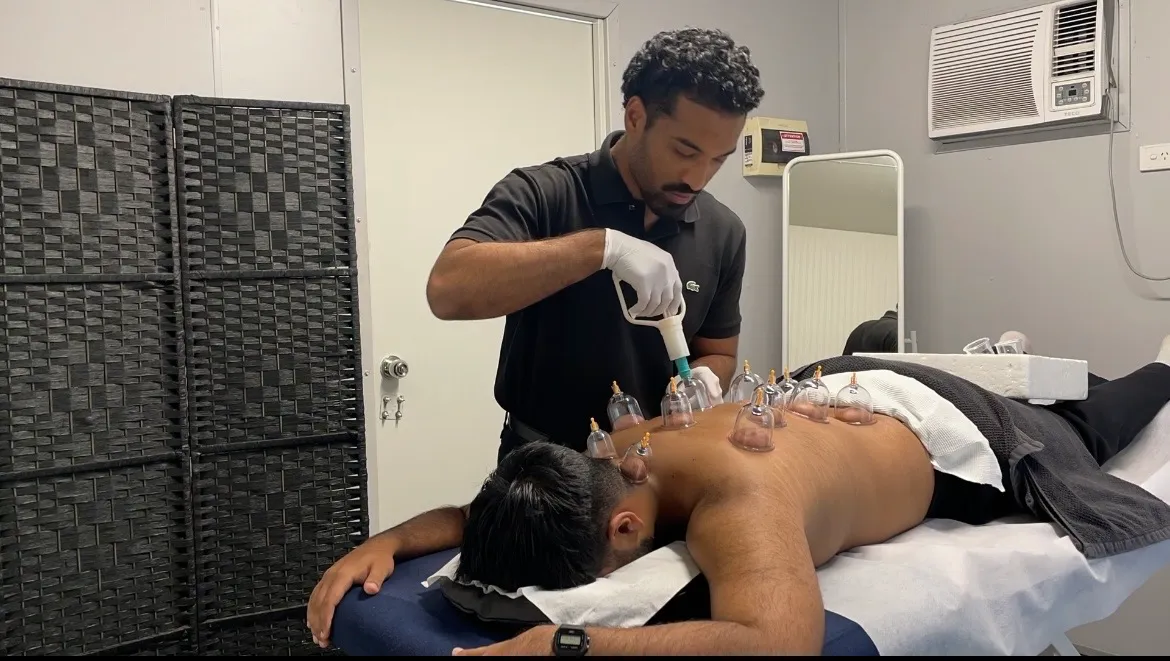
Where the cups go
Point selection is not mysterious and it is not improvised. It comes down to which of three situations you are in when you walk in.
-
1
General, or Sunnah, points
For a general session where there is no specific complaint. Five main points along the back, sitting over the major organs, of which the top three are the ones most practitioners recognise as the Sunnah points. For a lot of people that is the whole session, and it is the one most families already know.
-
2
Specific pain
For a targeted area: neck, shoulders, back or head. I find the spot by pressing around the region and going by what you tell me, then the cups go on and around it. That is why two people with sore shoulders do not necessarily get cups in the same places.
-
3
Lower abdomen and pelvis
A separate group of points used for conditions in that region, including sexual health complaints. This is a conversation before it is a decision, it is not part of a general session, and plenty of people never need it.
What decides it on the day
Screening first, then what you came in for. I go through your history, medications and anything your GP is managing before a single cup is opened, and if that rules Hijama out on the day there is no charge. Points are a conversation, not a chart I apply to you.
And what about the blood?
People look at what is in the cup and want to know what it means. It is the single most common question I get after a first session, usually phrased as some version of “so was there a lot of bad stuff in mine?”
The material people most often send me on this comes from David Parker ND, DIP ION, DIP EAV, who works in clinical nutrition, naturopathy and research into both modern and traditional practice. His approach is to assess blood under a microscope to read nutritional status and the elements he identifies in a sample, comparing a picture before a therapy with one after it.
By his own account he was, in his words, “intrigued to explore the scientific reasons” behind why cupping appeared to help across such a spread of complaints. Wet cupping interested him most, so he began examining blood drawn through the cups under both dark-field and light-field microscopy.
What he reports finding: dried layered samples showing, as he reads them, high concentrations of toxic metals and chemicals along with signs of bacterial and parasitic activity, and live samples under dark field showing acids and inflammatory proteins. He reports these appeared more frequently when the blood had been drawn from an area where the person was experiencing pain and inflammation.
From that he concludes the painful area “appears to act like a magnet for acids, toxins and pathogens”, and reasons that clearing them locally would bring symptomatic relief while fresh circulation delivers nutrients and oxygen. He regards wet cupping as a useful adjunct alongside dietary change and reducing what a person is exposed to in the first place.
The six pictures he describes
His write-up is built around six microscope images. In plain terms, this is what he says each one shows:
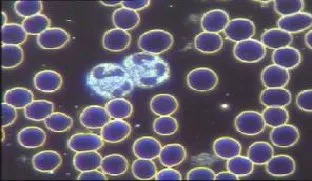
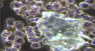
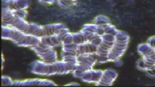
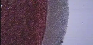
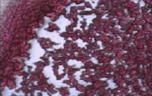
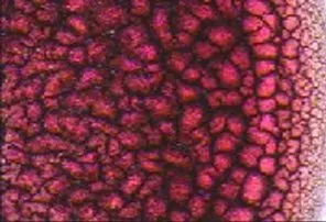
Microscope images and their interpretation: David Parker ND, DIP ION, DIP EAV, reproduced from his work. The wording above is my summary of what he states each image shows. They are not photographs of any client of mine, and they are not findings of mine.
Where I stand on it
I have set that out at length because I get asked, and because I would rather hand you the source and my view of it than a summary of a summary. Now the part that matters.
I find it genuinely interesting, and I think careful observation is how a lot of medicine has started. What it has not had yet is the kind of testing that would let anyone call it settled, and microscopy like this is not a diagnostic test. So I hold it the way I would hold any promising observation: worth looking at, not something to hang a promise on.
How I work with it in practice
- I keep microscopy out of the treatment room. It is a research interest, not part of what I offer you.
- I will happily talk through the toxin and pathogen theories with you. What I will not do is state them as fact, because a cup cannot tell us that either way.
- The colour and texture of what comes out varies for all sorts of ordinary reasons, so I do not read your health in it.
- If something is unexplained, it still needs proper investigation with your GP. Hijama sits alongside that, not instead of it.
What I can speak to confidently is what the cup does mechanically, which is drawn out layer by layer on the Hijama page, and what people actually report afterwards: lighter and looser through the area worked on, often calmer for a day or two, and sometimes a bit tired that evening.
Ask me the awkward questions first
If you have never had Hijama, call and ask before you book. I would rather spend ten minutes on the phone than have you turn up unsure.
This article is general information, not a diagnosis or personal health advice. Hijama is not a substitute for medical investigation or treatment. Keep taking prescribed medication and keep seeing your GP, and get anything unexplained looked at properly.